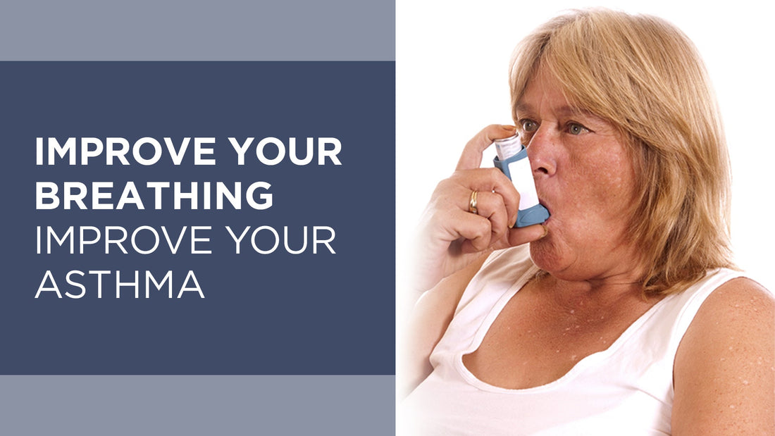
Improve Your Breathing - Improve Your Asthma
Asthma is a condition that restricts airflow due to narrowed airways, making breathing difficult. There has been a sharp increase in the global prevalence, morbidity, mortality, and economic burden associated with asthma over the last 60 years, particularly in children. Nearly 28 million people in the U.S are affected by asthma, according to the CDC (Centers for Disease Control and Prevention). This equals about 1 in 12 people. Approximately 260 million people worldwide currently have asthma.
The Connection Between Asthma and Breathing Habits
Research and anecdotal evidence suggest that poor breathing habits contribute to asthma symptoms, and improving these habits can serve as an effective complement or alternative to medication. However, mainstream healthcare has yet to fully embrace this approach. Despite this, logic dictates that breathing quality directly influences conditions affecting the airways. Many individuals have found significant relief through breath training, sometimes even reducing or eliminating their need for medication.
Breath Training for Asthma Management
Many asthma sufferers are unaware of how transformative proper breathing can be. Testimonials from individuals who have improved or even reversed their symptoms through Conscious Breathing practices are abundant.
For instance, Mattias Andersson, a 37-year-old former Nordic Taekwondo Champion, had struggled with asthma for years. After participating in a Conscious Breathing course, his symptoms drastically improved.
– I haven’t used asthma medication for nearly two months—it’s incredible! Thank you for a brilliant course, and particularly for a revolutionary discovery. Before the course, I took Bricanyl two to five times a day and Pulmicort twice daily. Since learning better breathing techniques, my symptoms have significantly diminished. I’m truly grateful—in a way you have changed my life.

What Is Asthma?
Asthma is characterized by difficulty breathing due to airway constriction, limiting airflow to and from the lungs. When we inhale, oxygen travels to the alveoli, where it enters the bloodstream, while carbon dioxide is expelled during exhalation.

The airways branch out 23 times before reaching the alveoli, narrowing progressively.
So the air travels quite a long distance to reach the alveoli. This distance can be compared to a snorkel. In this “snorkel,” nothing happens; it’s simply a transportation route. In medicine, this transportation route is called “the dead space.”
We have hundreds of millions of alveoli, and the majority are found in the lower part of the lungs. This is also where most of the blood flow, ten times as much as in the upper regions.
If airway diameter is reduced by 50%, 16 times more effort is required to move air in and out of the lungs.
In asthma, the airways are more narrow than normal, contributing to inefficient air transport. This inefficiency forces asthmatics to breathe more rapidly, leading to further complications.
When the airways are narrow, breathing becomes shallower, and less air reaches the alveoli and the blood. This is a very inefficient way to breathe, and we are forced to compensate by breathing faster.
Narrow airways are sort of like driving with the emergency brake slightly engaged. You get to where you need to be, but to reach the desired speed, you have to push the gas pedal harder. The most effective method, is obviously to disengage the brake, in other words, to find the cause of the narrow airways and solve it.
Common Asthma Symptoms
When an asthma attack occurs, the exhalation is particularly difficult, and a serious attack can be potentially lethal. The most common asthma symptoms are:
- Shortness of breath
- Wheezing
- Chest tightness
- Coughing
- Excessive mucus production
- Inflammation in the airways
- Nasal congestion
- Fatigue
- Sleep problems
- Allergies
- Anxiety and depression
How Asthma Medication Works
Asthma medications typically fall into two categories:
- Bronchodilators (similar to adrenaline) relax airway muscles, widening them for easier breathing.
- Corticosteroids (similar to cortisone) reduce inflammation in the airways.
While these medications provide symptom relief, they do not address the root causes. As soon as one stops the medication, the symptoms return immediately. Additionally, because both adrenaline and cortisol are stress hormones, prolonged use may contribute to heightened anxiety and difficulty relaxing.
Are you your own asthma diagnosis?

Anna Sundkvist Kräutner has been diagnosed with asthma. She works as a conversational therapist and has educated herself as a Conscious Breathing Instructor. Anna says:
– To not be able to breathe is terrible, anyone who’s experienced it knows that. The panic, stress, and anxiety are sky high. I myself have asthma and pollen allergies, but the problems are largely non-existent nowadays, after four years of breath training. This is something I am incredibly happy about. Many asthmatics/people with allergies feel like they are their condition and turn their entire body and their thoughts to ‘NOW the pollen season begins, and I will get sick.’ The body becomes tense, breathing speeds up.
Earlier, when I visited someone with a cat or a dog, my body and thoughts were already on the way there for having an asthma attack.
Now, I know that I can use my breathing to help if my airways, against all odds become narrow, and I am, in other words, entirely relaxed. Just the knowledge that I always have a powerful tool with me, no matter where I go, in breathing, automatically relaxes me.
I have taken the Conscious Breathing Instructor course and now I spread this important knowledge to my clients. When they understand how it all connects and realize that they can affect their situation on their own, many stop being victims of their diagnosis. Everyone with asthma and allergies should learn to breathe better.
Five Key Factors Affecting Asthma and Breathing

Many asthmatics hyperventilate, taking in too much oxygen and expelling too much carbon dioxide. Since CO2 helps relax airway muscles, low levels contribute to airway constriction. Studies confirm that asthmatics tend to have an elevated breathing rate, which exacerbates symptoms.
Narrow airways due to a shortage of carbon dioxide, can be viewed as a defense mechanism, which has the purpose of preventing the outflow of carbon dioxide, which almost exclusively leaves the body via exhalation, so that optimal levels are retained in the body.
When we breathe in a relaxed way, the breathing volume decreases, and the body can then normalize the CO2-pressure. The narrow airways then widen as the defense mechanism is no longer required.
Here are some scientific studies which confirm the correlation between over-breathing and asthma.
| Liters per minute | Participants | Year | Reference |
|---|---|---|---|
| 14.0 | 39 | 1998 | Buteyko breathing techniques in asthma: a blinded randomised controlled trial (Ref 1) |
| 13.4 | 17 | 1982 | Respiratory center output and ventilatory timing in patients with acute airway and alveolar (pneumonia) disease (Ref 2) |
| 13.3 | 16 | 2004 | Ultrafine particle deposition in subjects with asthma (Ref 3) |
| 12.0 | 101 | 1968 | Arterial-blood gas tension in asthma (Ref 4) |

The sinuses produce nitric oxide (NO), which helps relax and widen the airways while also acting as an antimicrobial agent. Nasal breathing ensures that NO travels with the inhaled air, helping to open the airways and improve airflow. Once in the lungs, NO enhances oxygen absorption by dilating blood vessels, facilitating more efficient oxygen transfer into the bloodstream.
In contrast, mouth breathing bypasses this process, depriving the lungs of NO’s beneficial effects. As a result, airway constriction may increase, making breathing less efficient and raising susceptibility to infections.

The body’s natural response to injury involves inflammation, which includes redness, heat, swelling, and pain—essential first steps in healing. For example, when you sprain your ankle, blood vessels expand to increase blood flow, boosting oxygenation and energy production. This process generates heat and carbon dioxide, while fluid accumulation causes swelling.
Pain intensifies as chemicals are released and nerves become stimulated. Swelling and pain serve as protective mechanisms, limiting movement to prevent further injury. Persistent pain and swelling indicate that healing is still in progress.
The inflammatory response functions similarly in the airways. Breathing in cold, dry air; airborne allergens like pollen; bacteria; viruses; pollution; or poor indoor air can trigger inflammation, causing the airways to swell. This swelling restricts airflow, leading to shallower breathing.
To compensate, we breathe faster, but this increases mechanical stress on the airways, further aggravating inflammation and creating a vicious cycle. If left unaddressed, inflammation may become chronic, potentially causing permanent structural damage, as seen in conditions like pulmonary emphysema and Chronic Obstructive Pulmonary Disease (COPD).


Researchers at Karolinska Institute in Sweden estimate that we inhale up to 100 billion particles daily, with even higher exposure in polluted urban environments.
Mucus production in the airways plays a crucial role in the body’s defense system, trapping and preventing harmful particles from reaching the lungs. For example, smokers inhale large amounts of foreign particles, prompting the body to produce excess mucus, leading to the characteristic “smoker’s cough.” Similarly, mucus production increases during inflammation, such as with a blocked nose.
While mucus helps neutralize bacteria and other harmful particles, excessive production can narrow the airways, making breathing more difficult. Prolonged overproduction can lead to permanently enlarged mucous glands, contributing to chronic airway constriction.
Mouth breathing bypasses the body's first line of defense—the nose. The nasal passages not only warm and humidify inhaled air but also filter out bacteria and particles before they reach the lungs. Additionally, exhaling through the nose re-warms and re-humidifies the nasal passages, which become cooler and drier after inhalation, and helps expel trapped particles, making it an efficient heat exchanger and self-cleaning filter.


Water is essential for keeping the airways moist and functioning properly. In fact, our lungs are made up of 80% water! With every breath, we lose water, as exhaled air is 100% humidified, whereas inhaled air rarely exceeds a humidity level of 70-80%. During dehydration, the body naturally constricts the airways to minimize further water loss.
This is yet another example of the body's built-in defense mechanisms. When we understand how our body works, we see that it is always striving to protect us. Our body is not our enemy—it is constantly working in our best interest.
A visible example of water loss during breathing occurs in cold weather, where exhaled air appears as vapor. This "winter breath" is a mix of heat and moisture. A Swedish study found that exhaling through the mouth results in 42% more water loss than exhaling through the nose. Nasal breathing helps conserve both heat and moisture, making it a more efficient way to breathe.
When the body lacks sufficient water, mucus production decreases, leaving the lungs more vulnerable to irritation. This can make individuals more susceptible to bacteria, viruses, and airborne particles like dust and mold.
For individuals with asthma, maintaining proper hydration and breathing through the nose is especially important. However, balance is key—drinking too much water can be just as problematic as drinking too little.
Improved Breathing Habits Can Relieve Asthma Symptoms
Poor breathing habits are common among asthmatics. In a study of 38 individuals with asthma, 95% were found to have ineffective breathing patterns. After receiving training in proper breathing techniques, 90% experienced a reduction in asthma symptoms. (Ref 5)
Three studies conducted in New Zealand further support these findings. The results showed a 66-95% decrease in the use of bronchodilators (adrenaline-like medication) and a 41-50% decrease in the use of corticosteroids (inflammation-suppressing cortison-like medication) among participants who improved their breathing. One study also noted that, in addition to reduced medication use, participants reported fewer symptoms and an improved quality of life. (Ref 6-8)
A growing body of research reinforces this connection, and many asthmatics who have adopted better breathing habits have been able to reduce or even discontinue their medication. There is no doubt that improving ones breathing habits can play a significant role in managing and alleviating asthma symptoms.
| Effect on medicine use | Brisbane | Gisborne | Gisborne children |
|---|---|---|---|
| Decrease of bronchodilators | 95% | 85% | 66% |
| Decrease of inflammation suppressors | 49% | 50% | 41% |
The Link Between Asthma, Depression, and Anxiety
A person who cannot use their legs may rely on a wheelchair, just as someone struggling to breathe depends on asthma medication. Given that most asthmatics hyperventilate, incorporating breath training into asthma treatment should be a standard approach.
Research has shown a strong connection between asthma, anxiety, and depression. A meta-analysis of surveys—including 83,000 personal interviews across 17 countries—found that anxiety, depression, and alcoholism were 50% more common in asthmatics compared to non-asthmatics. (Ref 9)
Dr. Alicia Meuret conducted an extensive study tracking over 100 individuals for several years, documenting their breathing patterns. Her research revealed that asthmatics, as well as those suffering from anxiety, tend to over-breathe, leading to lower carbon dioxide levels in the body.
The study also found that approximately an hour before an asthma or anxiety attack, participants’ breathing patterns became slightly more erratic. While the triggers varied, this subtle change was enough to further reduce already low carbon dioxide levels, increasing the likelihood of an attack.
Preventing An Asthma Attack
To prevent the panic response during an asthma attack, the body needs to relax—easier said than done, but entirely possible. A person experiencing an attack often begins to hyperventilate, leading to shortness of breath.
Exhalation is particularly difficult during an asthma attack, often accompanied by a high-pitched wheezing or rattling sound. This occurs because stress causes rapid and forceful exhalation, further tightening the airways.
A highly effective way to regain control is through low, slow, and rhythmic breathing. To achieve this, focus on prolonging the exhale to allow for a lower, more relaxed inhalation. This mirrors the natural response when danger has passed—exhaling with a sigh, lowering the shoulders, and relaxing the body.
One simple method to extend exhalation is to slightly constrict the throat, creating resistance while softly pushing the air out. Alternatively, using a breathing tool like the Relaxator can help regulate exhalation. The inhale should be low, slow, small, and rhythmic through the nose. This technique opens the airways naturally, reducing the need for stress hormones like cortisol (which reduces inflammation) and adrenaline (which widens the airways).
As carbon dioxide levels normalize through controlled breathing, muscle cramping in the airways subsides, allowing for easier breathing. Nasal inhalation further warms, humidifies, and filters the air from bacteria, reducing inflammation and minimizing excessive mucus production.
What can you do on your own?
- Get to know your asthma. To be able to prevent your asthma problems, you need to get to know them, and become aware of which environments, situations, and foods trigger or increase your asthma symptoms. Avoid foods that help form mucus and give rise to inflammation, for example, sugar, wheat, and dairy. Make sure to drink enough water.
- Improve your breathing. Figure out where your breathing is by answering the questions in The Breathing and Health Index. If the results show that your breathing habits have room for improvement, you will likely benefit from doing The 28-day Conscious Breathing Retraining Program.
- Unblock a blocked nose. If your nose feels tight and you struggle to breathe through it, it is often a sign of impaired breathing. In your nose, under the turbinates, there are erectile tissues. As you improve your breathing, they will decrease in size and your nasal passages will become less tight. Learn more about how to unblock a stuffy nose.
- Close your mouth. We often catch colds during the night, and one reason is that we sleep with our mouths open, which cools down our nose. To tape your mouth shut at night with Sleep Tape is an extremely easy, yet very powerful tool. Since it isn’t easy to watch your breathing while you sleep, the use of Sleep Tape guarantees that your mouth remains closed at night so your breathing only occurs through the nose.
- Breathing retraining. Retrain your breathing with the Relaxator, or by pursing your throat a little to create a resistance on the exhale, and then softly push out the air. This prolongs the exhalation, which creates a slower but more relaxed breathing and allows for a lower inhalation. Additionally, you strengthen your immune system by doing low-intensity exercise with a closed mouth.
This article is based on the book Conscious Breathing. Thank you for taking the time to read this, I hope you enjoyed it.
Scientific studies
Title: Buteyko breathing techniques in asthma: a blinded randomised controlled trial
Authors: Bowler SD1, Green A, Mitchell CA
Journal: Med J Aust. 1998 Dec 7-21;169(11-12):575-8
Abstract:
OBJECTIVE: To evaluate the effect of Buteyko breathing techniques (BBT) in the management of asthma.
DESIGN: Prospective, blinded, randomised study comparing the effect of BBT with control classes in 39 subjects with asthma. The study was conducted from January 1995 to April 1995.
PARTICIPANTS AND SETTING: Subjects recruited from the community, aged 12 to 70 years, with asthma and substantial medication use.
MAIN OUTCOME MEASURES: Medication use; morning peak expiratory flow (PEF); forced expiratory volume in one second (FEV1); end-tidal (ET) CO2; resting minute volume (MV); and quality of life (QOL) score, measured at three months.
RESULTS: No change in daily PEF or FEV1 was noted in either group. At three months, the BBT group had a median reduction in daily beta 2-agonist dose of 904 micrograms (range, 29 micrograms to 3129 micrograms), whereas the control group had a median reduction of 57 micrograms (range, -2343 micrograms to 1143 micrograms) (P = 0.002). Daily inhaled steroid dose fell 49% (range, -100% to 150%) for the BBT group and 0 (range, -82% to +100%) for the control group (P = 0.06). A trend towards greater improvement in QOL score was noted for BBT subjects (P = 0.09). Initial MV was high and similar in both groups; by three months, MV was lower in the BBT group than in the control group (P = 0.004). ET CO2 was low in both groups and did not change with treatment.
CONCLUSION: Those practising BBT reduced hyperventilation and their use of beta 2-agonists. A trend toward reduced inhaled steroid use and better quality of life was observed in these patients without objective changes in measures of airway calibre.
Title: Respiratory center output and ventilatory timing in patients with acute airway and alveolar (pneumonia) disease
Authors: Kassabian J, Miller KD, Lavietes MH
Journal: Chest. 1982 May;81(5):536-43
Abstract: To investigate the mechanism for hyperventilation in two common but dissimilar conditions, asthma (A) and lobar pneumonia (P), we examined the ventilatory pattern in 17 A and six P subjects. When acutely ill, hyperventilation (PaCO2) was similar in the two groups. Minute ventilation (VE) however, was slightly greater in group A than in P. In A patients, measurements of occlusion pressure (dP/dT) and inspiratory flow rate (VT/ti) during quiet breathing showed enhancement of respiratory center output. By contrast, in P patients, dP/dT and VT/ti were not elevated. Tidal volume (VT) was 0.59 +/- 0.19 L in A; 0.45 +/- 0.09 in P. Respiratory rate was increased in both groups. With supplementary oxygen therapy, neither VE nor PaCO2 changed in either group. The mechanism for the increased ventilatory drive in group A is unclear. Most likely, reflexes initiated by either bronchospasm or by the sudden increase of end-expiratory lung volume (EELV), not operative in P, account for the increased respiratory center output seen in A. To examine the latter possibility, we studied the ventilatory pattern at both normal and increased EELV in six nonasthmatic subjects. Both dP/dT and VT/ti were increased in all subjects after elevation of EELV. Thus, changes in EELV may be important for the regulation of ventilation during bronchospasm.
Title: Ultrafine particle deposition in subjects with asthma.
Authors: Chalupa DC1, Morrow PE, Oberdörster G, Utell MJ, Frampton MW
Journal: Environ Health Perspect. 2004 Jun;112(8):879-82
Link to full text: Access here 
Abstract: Ambient air particles in the ultrafine size range (diameter < 100 nm) may contribute to the health effects of particulate matter. However, there are few data on ultrafine particle deposition during spontaneous breathing, and none in people with asthma. Sixteen subjects with mild to moderate asthma were exposed for 2 hr, by mouthpiece, to ultrafine carbon particles with a count median diameter (CMD) of 23 nm and a geometric standard deviation of 1.6. Deposition was measured during spontaneous breathing at rest (minute ventilation, 13.3 +/- 2.0 L/min) and exercise (minute ventilation, 41.9 +/- 9.0 L/min). The mean +/- SD fractional deposition was 0.76 +/- 0.05 by particle number and 0.69 +/- 0.07 by particle mass concentration. The number deposition fraction increased as particle size decreased, reaching 0.84 +/- 0.03 for the smallest particles (midpoint CMD = 8.7 nm). No differences between sexes were observed. The deposition fraction increased during exercise to 0.86 +/- 0.04 and 0.79 +/- 0.05 by particle number and mass concentration, respectively, and reached 0.93 +/- 0.02 for the smallest particles. Experimental deposition data exceeded model predictions during exercise. The deposition at rest was greater in these subjects with asthma than in previously studied healthy subjects (0.76 +/- 0.05 vs. 0.65 +/- 0.10, p < 0.001). The efficient respiratory deposition of ultrafine particles increases further in subjects with asthma. Key words: air pollution, asthma, deposition, dosimetry, inhalation, ultrafine particles.
Title: Arterial-blood gas tension in asthma
Authors: McFadden ER Jr, Lyons HA
Journal: N Engl J Med. 1968 May 9;278(19):1027-32
Abstract: Arterial oxygen and carbon dioxide tensions, pH and forced expiratory volumes were measured in 101 asthmatic patients during acute attacks of bronchospasm. Hypoxemia was observed in 91 subjects. The cause of the depressed oxygen tensions was found to be an alteration in ventilation-perfusion ratios. Overall alveolar hypoventilation and increased venous admixture were found to contribute to the hypoxemia in some patients with very severe levels of airway obstruction. Seventy-three subjects had hypocarbia and respiratory alkalosis. Carbon dioxide retention (observed in 11 patients) occurred only at extreme degrees of obstruction. Age, history of asthma and duration of the acute attack were unrelated to the alterations in blood gas tensions, pH or severity of airway obstruction.
Title: Regulation of ventilatory capacity during exercise in asthmatics
Authors: Johnson BD, Scanlon PD, Beck KC
Journal: J Appl Physiol (1985). 1995 Sep;79(3):892-901
Abstract: In asthmatic and control subjects, we examined the changes in ventilatory capacity (VECap), end-expiratory lung volume (EELV), and degree of flow limitation during three types of exercise: 1) incremental, 2) constant load (50% of maximal exercise capacity; 36 min), and 3) interval (alternating between 60 and 40% of maximal exercise capacity; 6-min workloads for 36 min). The VECap and degree of flow limitation at rest and during the various stages of exercise were estimated by aligning the tidal breathing flow-volume (F-V) loops within the maximal expiratory F-V (MEFV) envelope using the measured EELV. In contrast to more usual estimates of VECap (i.e., maximal voluntary ventilation and forced expiratory volume in 1 s x 40), the calculated VECap depended on the existing bronchomotor tone, the lung volume at which the subjects breathed (i.e., EELV), and the tidal volume. During interval and constant-load exercise, asthmatic subjects experienced reduced ventilatory reserve, higher degrees of flow limitation, and had higher EELVs compared with nonasthmatic subjects. During interval exercise, the VECap of the asthmatic subjects increased and decreased with variations in minute ventilation, due in part to alterations in their MEFV curve as exercise intensity varied between 60 and 49% of maximal capacity. In conclusion, asthmatic subjects have a more variable VECap and reduced ventilatory reserve during exercise compared with nonasthmatic subjects. The variations in VECap are due in part to a more labile MEFV curve secondary to changes in bronchomotor tone. Asthmatics defend VECap and minimize flow limitation by increasing EELV.
Title: Hyperventilation – Asthma
Authors: Herxheimer H
Journal: The Lancet. 1946 Jan 19;83-86
Abstract: Bronchial asthma is a syndrome rather than a disease and can be brought on by allergic factors outside the patient (extrinsic), or within the patient (intrinsic), or both. The intrinsic may be metabolic (endocrine), psychological (“nervous asthma “), infection (acute bronchitis), or primary emphysema. In all cases bronchial spasm followed by asthma is, elicited very easily, and in many patients, once the syndrome has been established, the spasm can be elicited by various ” trigger ” factors, not only allergic but also of different origin.
It seems to be certain that this disposition to bronchial spasm can be present from birth-many children develop asthma very early-but there is an important group of intrinsic asthma where the syndrome develops only at the age of about 40, thus suggesting that it is acquired. Even in these cases the tendency may have been present from birth-the gun may have been loaded, to use the metaphor of Rackemann (1938), but the trigger has not been pressed till late, when a suitable trigger factor developed. One of these trigger mechanisms to which little attention seems to have been paid is hyperventilation and in some cases hyperventilation seems to be the only cause of the asthma.
It is difficult to estimate in what proportion of cases of asthma hyperventilation through excitement or muscular exercise acts as a releasing mechanism. In many cases voluntary hyperventilation has no such effect, and in others muscular exercise is well tolerated. In some of them voluntary hyperventilation even seems to be beneficial.
Title: Controlling Asthma by Training of Capnometry-Assisted Hypoventilation (CATCH) vs Slow Breathing
Authors: Alicia E. Meuret et al.
Journal: Chest. 2014 Nov; 146(5): 1237–1247.
Link to full text: Access here
Abstract:
BACKGROUND: Hyperventilation has been associated with adverse effects on lung function, symptoms, and well-being in asthma. We examined whether raising end-tidal CO2 levels (ie, Pco2) compared with slow breathing is associated with improvements in asthma control, including peak flow variability.
METHODS: One hundred twenty patients with asthma were randomly assigned to capnometry-assisted respiratory training (CART) for raising Pco2 or slow breathing and awareness training (SLOW) for slowing respiratory rate. Patients received five weekly sessions and completed bid homework exercises over 4 weeks. Blinded assessments at baseline, posttreatment, 1- and 6-month follow-up of asthma control, Pco2, and diurnal peak flow variability were primary outcome measures. Additionally, we measured pulmonary function (spirometry, forced oscillation, exhaled nitric oxide, and methacholine challenge), symptoms, quality of life, and bronchodilator use. Because the control group received active treatment, we expected improvements in asthma control in both groups but more pronounced benefits from CART.
RESULTS: Improvements were seen in 17 of 21 clinical indexes (81.0%) in both interventions, including the primary outcome variables asthma control (d = 0.81), peak flow variability (d = 0.54), quality of life, bronchodilator use, lung function, and airway hyperreactivity. Most improvements were sustained across the 6-month follow-up. Compared with slow breathing, CART showed greater increases in Pco2 (d = 1.45 vs 0.64 for CART vs SLOW, respectively) and greater reductions in respiratory impedance during treatment, less distress during methacholine challenge, and greater reduction in asthma symptoms at follow-up (P < .05).
CONCLUSIONS: Brief interventions aimed at raising Pco2 or slowing respiratory rate provide significant, sustained, and clinically meaningful improvements in asthma control. Raising Pco2 was associated with greater benefits in aspects of lung function and long-term symptoms.